Rhodesian Ridgeback
HISTORY of the BREED
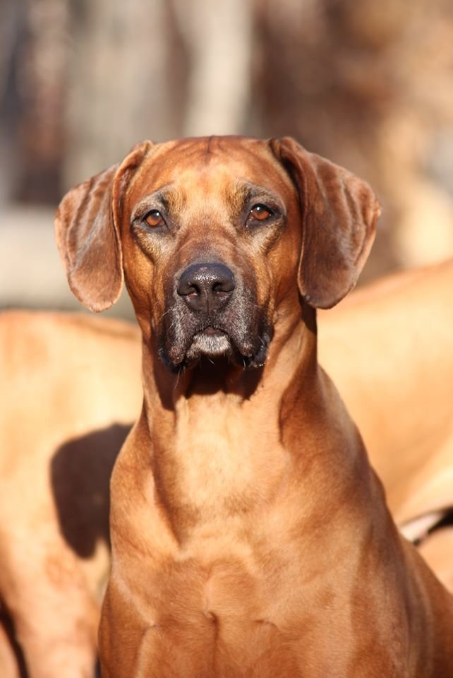
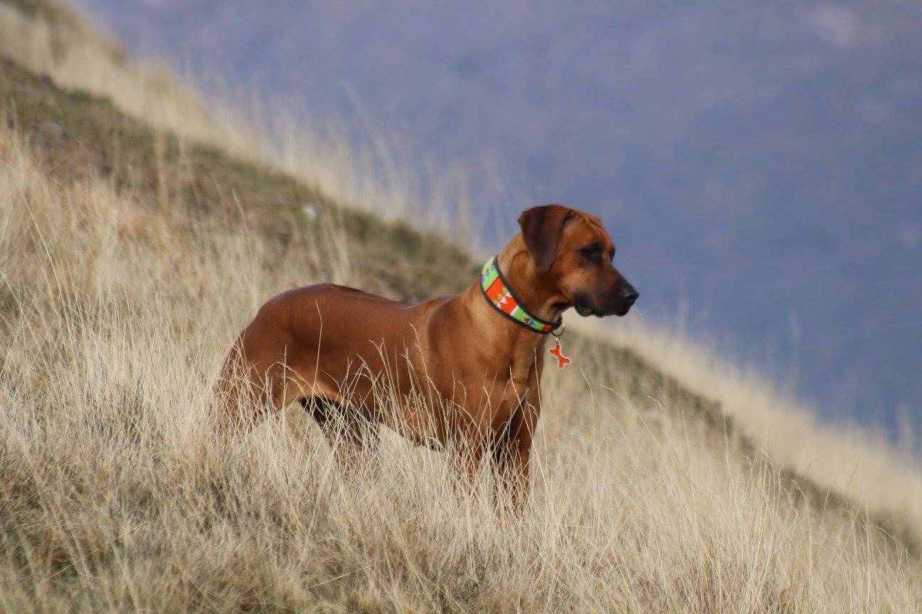
The Rhodesian Ridgeback is the only recognized breed originating in Southern Africa and is one of the two recognized breeds that have on their back the typical fur crest that grows in the caudocardinal direction, a characteristic from which it also takes its name (ridge = crest, back = back ).
The RR is a breed whose history is intertwined with the history of Africa, of some of its tribes and of the first European settlers who landed in the Cape province in the mid-1600s. The following article, written by a well-known American breeder, Mrs. Sawyer , summarizes the concept of Ridgeback very cleverly.
As Ms Sawyer says, the RR evolved in Rhodesia, today's Zimbabwe, on the basis of a breed previously created, but not recognized, in South Africa: the "Boer dog". But the origin of the dogs with the mane is certainly more ancient: in Africa the crest appears very often in the most isolated villages, and is also found in Asia. The best known example is the dog on the island of Phu Quoc, off Thailand; if there is a connection between this Asian dog and the dog with the African mane, it could go back to the various trade between Asia and Africa, starting with those of the Phoenicians, on the North African coasts, or of the Arabs, on the Eastern coasts, or more recently of the Portuguese or of the Dutch on the Mendenian coasts. But it could also be more simply about parallel mutations and, however, in Africa, the crest takes us back to an indigenous dog, the Khoi Khoi, or "Hottentot dog", of which we find the first representation in the book by Dr. David Livingstone from 1870 entitled Livingstone's Missionary Travels in South Africa, the first descriptions by some historians of the 1700s (Kolben, Theal, etc.) and a mysterious painting on rock in Zimbabwe, where the dog with the mane is depicted among the great Hottentot shepherds and their cattle, the Zebu vaccines and the Sori sheep, ancestors of today's South African cattle.
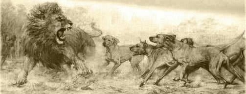
 With the Hottentots, the Zebu vaccines and the Sori sheep even the dog with the mane migrated, from 500 to 1500, across Africa, from the coasts of the Red Sea to the Cape of Good Hope where, in 1600, with the advent of first Europeans, its transformation began. Used successfully by settlers for hunting and defense, it was crossed with the breeds then present in the region, especially Pointer, Deerhound or Greyhound, and Bull Terrier. The resulting "steekbaar", also called "Boer dog", often had a mane on his back and, always, this was synonymous with particular qualities of adaptability to the so varied climates of South Africa; qualities of faithful and courageous guardian, endowed with a great instinct for hunting, in which the primitive and rough Khoi Khoi was refined thanks to the introduction of blood from the most refined European breeds. While the selection was based only on principles of utility that imposed only the survival of the fittest, the colonial type of life and perhaps even the vastness of the country did not give way to a strictly dog-like awareness and activity, so much so that the morphological characteristics they were never established and, with the rapid change of living conditions, the race ran the risk of getting lost. But in 1870 the English Reverend Charles Helm brought a pair of Boer dogs to his new mission in Matabeleand, Rhodesia: like the Boers 200 years earlier, so then the settlers of this African region were conquered by the qualities of this dog built to measure for African colonial life and, with the further introduction of blood Collie and Airdale, they obtained a powerful, but not heavy, dog with particular agility and endurance and incredible courage, tempered by an instinctive respect for large African prey. Thus it was that the "Boer dog" became the "lion dog", used by the great hunters of Africa such as Selous' van Rooyen and Upcher.
 In 1922 Francis R. Barnes founded the Rhodesian Ridgeback (Lion Dog) Club "in Bulawayo, Rhodesia", today "Parent Club" and in 1924 he set the breed standard, approved the same year by the then South African Kennel Union. It was only after the Second World War that the RR regained the popularity of the "Boer dog" in South Africa: in 1945 the South African RR Club was founded and in 1982 the RR Club Transvaal. The Ridgeback, created both as a hunting dog and as a guardian and faithful companion of man, of an almost exclusively wolf-like character, and therefore it is not easy and understandable for everyone; tenacious and independent, he "possesses" only one master, capable of an almost degrading sweetness with children, unfriendly with strangers and becomes a proud guardian in front of the intruder, however always dominated by the balance necessary to make him a trusted friend. In many countries it is used for hunting: wild boars and goats in Australia, cats and deer in North America, large an tilopes and warthogs in Africa; to underline that at the same time as the hunter, the Ridgeback is always used also as a guardian, a job in which, even at a competitive level, it stands out for its instinctive qualities.
The Ridgeback is not only a beautiful dog to look at for the harmony of its forms, but a dog with a strong, very balanced character, "faithful and devoted to the master" as his ancestors were with the Hottentots, and like Boers and English they had conceived.
 From "I nostri cani" article by Giovanna Bacchini Carr (Breeder of "delle Cime Bianche")
STANDARD
ORIGIN : Southern Africa. Standard supplied by the Kennel Union of Southern Africa and the Zimbabwe Kennel Club.
DATE OF PUBLICATION OF THE OFFICIAL VALID STANDARD : 10.12.1996.
UTILIZATION : The Rhodesian Ridgeback is still used to hunt game in many parts of the world, but is especially prized as watchdog and family pet.
FCI-CLASSIFICATION : Group 6 Scenthounds and related breeds. Section 3 Related breeds. Without working trial. BRIEF HISTORICAL SUMMARY : The Rhodesian Ridgeback is presently the only registered breed indigenous to southern Africa. Its forbears can be traced to the Cape Colony of Southern Africa, where they crossed with the early pioneer’s dogs and the semidomesticated, ridged Hottentot hunting dogs. Hunting mainly in groups of two or three, the original function of the Rhodesian Ridgeback or Lion dog was to track game, especially lion, and, with great agility, keep it at bay until the arrival of the hunter. The original standard, which was drafted by F.R.Barnes, in Bulawayo, Rhodesia, in 1922, was based on that of the Dalmatian and was approved by the South African Kennel Union in 1926.
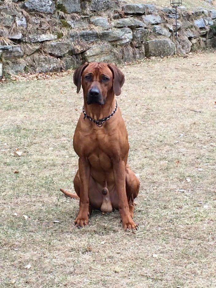
GENERAL APPEARANCE : The Rhodesian Ridgeback should represent a well balanced, strong, muscular, agile and active dog, symmetrical in outline, and capable of great endurance with a fair amount of speed. The emphasis is on agility, elegance and soundness with no tendency towards massiveness. The peculiarity of the breed is the ridge on the back, which is formed by the hair growing in the opposite direction to the rest of the coat. The ridge is the escutcheon of the breed.
 The ridge must be clearly defined, symmetrical and tapering towards the haunch. It must start immediately behind the shoulders and continue to the hip (haunches) bones. The ridge must contain only two crowns, identical and opposite each other. The lower edges of the crowns must not extend further down the ridge than one-third of its length. A good average width of the ridge is 5cm (2”).
BEHAVIOUR / TEMPERAMENT : Dignified, intelligent, aloof with strangers, but showing no aggression or shyness.
HEAD
CRANIAL REGION : Skull : Should be of a hair length (width of head between ears, distance from occiput to stop, stop to end of nose, should be equal), flat and broad between the ears; the head should be free from wrinkles when in repose. Stop : The stop should be reasonably well defined and not in one straight line from the nose to the occipital bone.
FACIAL REGION : Nose : The nose should be black or brown. A black nose should be accompanied by dark eyes, a brown nose by amber eyes. Muzzle : The muzzle should be long, deep and powerful. Lips : The lips should be clean, closely fitting the jaws. Jaws/Teeth : Jaws strong, with a perfect and complete scissor bite, i.e. the upper teeth closely overlapping the lower teeth and set square to the jaws. The teeth must be well developed, especially the canines or holders. Cheeks : Cheeks should be clean. Eyes : Should be moderately well apart, round, bright and sparkling, with intelligent expression, their colour harmonising with the colour of the coat. Ears : Should be set rather high, of medium size, rather wide at base, and gradually tapering to a rounded point. They should be carried close to the head.
 NECK : Should be fairly long, strong and free from throatiness.
BODY : Back : Powerful. Loins: Strong, muscular and slightly arched. Chest : Should not be too wide, but very deep and capacious; the brisket should reach to the elbow. Forechest should be visible when viewed from the side. Ribs moderately well sprung, never rounded like barrel-hoops. TAIL : Should be strong at the root and gradually tapering towards the end, free form coarseness. It should be of moderate length. It should not be attached too high nor too low, and should be carried with a slight curve upwards, never curled.
LIMBS
FOREQUARTERS : General appearance : The forelegs should be perfectly straight, strong and well boned, with the elbows close to the body. When viewed from the side, the forelegs should be wider than viewed from the front. Shoulders : The shoulders should be sloping, clean and muscular. Pastern : Should be strong with light spring.
HINDQUARTERS : General appearance : In the hind legs the muscles should be clean and well defined. Stifle : Good turn of stifle. Hock : Strong, well let down.
FEET : The feet should be compact and round, with well arched toes and tough, elastic pads, protected by hair between the toes and pads.
GAIT / MOVEMENT : Straight forward, free and active.
 COAT
COATHAIR : Should be short and dense, sleek and glossy in appearance, but neither woolly nor silky.
COLOUR : Light wheaten to red wheaten. A little white on the chest and toes is permissible, but excessive white hairs here, on belly, or above toes is undesirable. A dark muzzle and ears permissible. Excessive black hairs throughout the coat are highly undesirable.
SIZE AND WEIGHT : Height at withers : Dogs : 63-69 cm (25” -27”). Bitches : 61-66 cm (24” -26”). Weight : Dogs : 36,5 kg (80 lbs). Bitches : 32 kg (70 lbs).
FAULTS : Any departure from the foregoing points should be considered a fault and the seriousness with which the fault should be regarded should be in exact proportion to its degree and its effect upon the health and welfare of the dog.
DISQUALIFYING FAULTS: • Aggressive or overly shy dogs. • Any dog clearly showing physical of behavioural abnormalities shall be disqualified.

N.B.: • Male animals should have two apparently normal testicles fully descended into the scrotum. • Only functionally and clinically healthy dogs, with breed typical conformation should be used for breeding.
DISEASES
DERMOID BREAST IN THE RHODESIAN RIDGEBACK:
The dermoid sinus is a congenital malformation caused by a defect in the separation of the ectodermal embryonic leaflet from the neural tube, which is a portion of the same; from the ectodermal leaflet, during embryogenesis, the skin with its annexes (hair, sweat and sebaceous glands), and the nervous system originate.
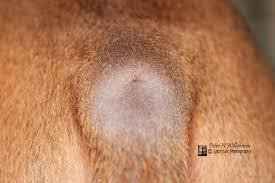
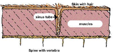
Its name is due to the particular shape: it is, in fact, a tubular invagination of the epidermis that extends from the surface of the skin, along the dorsal midline of the body, in the underlying tissues, up to a variable depth. It can be compared to a blind-bottomed sack that opens at the skin surface.
The internal walls of the sac are covered with skin with its annexes: hairs, sebaceous glands and atrophied sweat glands. The lumen of the breast is occupied by hairs and, with the passage of time, desquamative skin cells and sebum accumulate there, which are difficult to hesitate outside due to the reduced caliber of the lumen and the hairs that tend to retain the material that thus becomes an excellent medium for the growth of bacteria. In the past it has been called in various ways, among which hair cysts and African cysts; today the denomination of dermoid sinus is accepted which is classified as a particular type of dermoid cyst. The latter is defined as a roundish or oval swelling, localized deep in the dermis, of soft elastic consistency, of variable dimensions and covered by normal skin.
 Generally it does not open on the skin unless it becomes infected and gives rise to a fistula; it can be observed in all regions of the body, usually at the junction of the skin and subcutaneous tissues. The wall of the cyst is made up of skin, while the content is mainly made up of hair mixed with sebaceous material; it can also contain cartilaginous bone tissue and teeth. The dermoid sinus differs from the dermoid cyst proper in that it is located in the dermis and subcutaneous tissue, but can also extend to the underlying layers and opens onto the skin surface by means of an ostia from which they can protrude hair. As already mentioned, it is found exclusively along the dorsal midline both at the level of the head, both the neck and the trunk and can take on a cystic appearance only if it becomes infected.
The two congenital malformations also differ in the ways in which they are formed at the embryonic level: it now seems clear that the breast is formed following an incomplete fusion of the margins of the neural shower, from which the neural tube originates, along the midline of the embryo during the phases that lead to the formation of the nervous system on one side and to the structures from which the skin and its annexes will originate on the other. The dermoid cyst p.d. instead it seems to originate from the persistence of undifferentiated cells in the thickness of the subcutaneous connective tissues or in other sites which then "differentiate" in the skin tissue; according to other theories, it could derive from anomalies of formation of the skin appendages.
 In the past, the dermoid sinus has also been considered the equivalent of the human pathology known as the sinus or pilonidal fistula. This looks like a "dimple" located in correspondence of the coccygeal vertebrae, in which hairs are found which, unlike what is observed in the malformation of the dog, have the tip facing the inside of the breast. It is, in fact, a phlogistic lesion of a granulomatous type from a foreign body of the skin or subcutaneous tissue where foreign bodies are represented by free body hair of the same subject. It is therefore an acquired pathology, not present at birth, although in the past it was considered hereditary in nature most likely due to the fact that it may have a certain family predisposition for factors of physical constitution. The dermoid sinus is considered a malformation typical of the Rhodesian Ridgeback but occasionally it has also been found in dogs of other breeds: the scientific literature reports, in fact, two cases in the boxer, one case in the shih ztu one case in a boerboel and one case in a yorkshire terrier.
As for the location, in the Rhodesian Ridgebacks it seems that the breast was never found in the area occupied by the ridge, but only at the front or behind it.
The breast is found most frequently at the posterior part of the neck, in correspondence with the last cervical vertebrae, and in the interscapular region in front of the crest. Less frequently it is found at the level of the front of the cervical region of the neck, in correspondence with the second cervical vertebra, and of the sacral region in the space between the end of the crest and the base of the tail. In the same animal several dermoid sinuses can be found simultaneously, isolated or tending to flow.
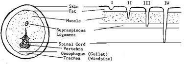
 There are four different types of dermoid sinus according to the classification of Lord and Mann & Stratton, which is based on its extension in depth: • Type I: the breast extends from the skin to the supraspinous ligament or the nuchal ligament;
• Type II: the breast does not reach these structures but is joined to them by a full fibrous connective tissue cord;
• Type III: it is similar to the previous ones only that it is not attached to the supraspinous ligament or to the nuchal ligament, but ends blindly at the level of the muscle tissue or subcutaneous tissue of the regions concerned (it is the most superficial).
• Type IV: it is the rarest and is most frequently detected at the level of the sacral region. The sinus ends at the level of the vertebral column and often connects to the dura mater, the outermost of the spinal meninges, running for a more or less long stretch in the vertebral canal.
 It can be said that this type of malformation, as well as congenital, is also hereditary in Rhodesian Ridgeback, unlike in other breeds. To date, the type of transmission is still not clear: at first it was hypothesized to be an autosomal recessive gene; subsequently the scholars turned towards a transmission linked to an autosomal dominant gene with incomplete penetrance. The most recent theories seem to again give credit to the recessive gene hypothesis. As a consequence of the fact that the exact transmission mechanism of the dermoid sinus has not yet been definitively clarified, it is difficult to eradicate the gene responsible for the malformation by selection. It is only possible to limit their diffusion by avoiding the use of dogs with dermoid breasts and, if necessary, also the breeders who, although not presenting the malformation, have produced more litters with dermoid breasts.
Specifying that the dermoid cyst cannot be considered a hereditary malformation based on the data provided by the scientific literature, the Rhodesian Ridgebacks that present this anomaly outside the typical localization areas of the dermoid sinus, should not necessarily be considered carriers of the gene responsible for the latter.
On the basis of the studies carried out in human medicine, a noticeable decrease in the incidence of the dermoid sinus has been found in puppies whose mothers before or in any case during the whole gestation period took a diet integrated with folate. However, it must be clarified that, according to current knowledge on the dermoid sinus, the use of folic acid can only decrease the percentage of puppies that show phenotypically the character, but cannot limit the hereditary transmission of the gene; this result is obtained only through a strict selection of the reproducers.
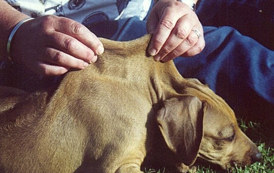
 Puppies with dermoid breasts at birth generally do not exhibit any symptoms. To highlight this malformation, the top of the back, neck and, if desired, of the occipital region can also be carefully examined, to try to highlight a small darker dot of the coat. from the hair that comes out of the breast. Often, however, it is not possible to appreciate the visual examination and in any case, to be certain, one must also resort to palpation which must be carried out, with great delicacy, on all Rhodesian puppies.
If the owner suspects, even vaguely, the presence of the breast, it is advisable to bring the puppies to the veterinary surgeon.
The dermoid sinus is a malformation that can be resolved surgically even if it is a type IV breast; in this case, however, it is necessary to attack the vertebral column.
It is considered appropriate to perform the intervention on the puppies early, in order to avoid that the breast can become infected, which leads to a wider tissue excision, which in turn determines the formation of disfiguring scars. If the operation is performed correctly, there are no clearly visible scars (except for complications), there are no recurrences, and the subjects can lead a completely normal life.
On the basis of what has just been stated, the use of euthanasia in all subjects presenting serious dermoid does not seem justified; however, it is clear that the use of the same as reproducers must be absolutely avoided, informing future owners of the consequences and possibly adopting systems to identify the operated subjects (tattoos for example), or resorting to their sterilization.
DYSPLASIA OF THE HIP
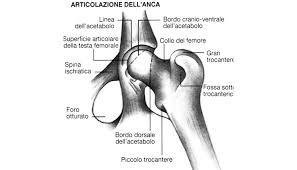
Hip dysplasia, from the Greek dys (abnormal) and plassein (formal), is an anomaly in the development of the coxo-femoral joint of the medium-sized dog, influenced not only genetically but also by multifactorial conditions, which results in algia dell hip and in secondary joint diseases. To date, new studies establish a genetic incidence of hip dysplasia at around 30%. The remaining 70% is of environmental, food and management origin.
 Pathogenesis
The critical period in the development of the dog's coxo-femoral joint goes from birth to 2 months of age: the bone tissue has not yet fully formed starting from the cartilaginous marrow, muscles and nerves are not yet fully developed and the soft tissues hip, still plastic, can be stressed beyond their limits of elasticity; this results in joint instability with loss of congruity between the acetabulum and the femoral head, subluxation of the femoral head, erosion of the articular cartilage, thickening of the synovial, appearance of exophytic formation, bone remodeling and osteoarthritis in the most advanced stages of hip dysplasia.
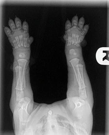
The loss of cohesion of the cartilage with the articular surface can occur as early as 60 days of age; joint laxity leads to a cranio-dorsal dislocation of the femoral head with a consequent greater load on the medial portion of the head and on the cranio-medial margin of the acetabulum. This abnormal pressure, in a 1-2 month old and already heavy puppy, causes a delay in the normal development of the acetabulum, with rounded edges. The articular surfaces undergo sclerosis in an attempt to stabilize the hip; there is also a thickening of the neck of the femur, the formation of osteochondrophytes on the same and subchondral bone cysts.
SYMPTOMATOLOGY
The onset and expression of clinical signs vary considerably in patients with hip dysplasia. The subject's reluctance to movement is frequently observed in an attempt to safeguard the sore hip. Other clinical signs are the modification of the running modality with a movement that decreases the joint excursion (running from the hare), difficulty in the passage from the decubitus to the station, reluctance to change position, pain to the manipulation of the coxo-femoral joint. Some subjects may exhibit unexpected changes in character by transforming themselves from balanced, sociable and game-oriented dogs into easily biting subjects.
The degree of lameness can vary from moderate (lameness only after very strenuous exercise) to extremely serious in which the dog is unable to maintain the quadrupedal station.
There are a number of factors that influence the manifestation of clinical signs, the most important of which is the age of the dog. Few puppies show pain in the first months of life even if they become severely affected by dysplasia during development.
The pain can appear suddenly and become evident at the palpitation of the hind limbs in puppies of 5-6 months, after jumping or climbing; sometimes there is difficulty in getting up especially from slippery floors.
Most dogs with dysplasia, after completing the first phase of abnormal development of the hip, walk and run without difficulty and without evident signs of pain up to 11-15 months of age, during which the pain appears only after intense activity and changes in gait become evident.
In adult dogs, pain is associated with arthritic phenomena; there is a tendency to sit up, atrophic muscle masses, stiffness in gait, shortened gait, signs that from intermittent become constant after 3-4 years.
The amount of pain is directly dependent on the size of the dog and its weight; training and training can mask asymptomatic hip dysplasia until adulthood until trauma or excessive physical activity occurs.
JUVENILE MYOCLONIC EPILEPSY (JME)
Juvenile Myoclonic Epilepsy Rhodesian Ridgeback Type (JME) is an inherited disorder characterized by myoclonic jerks and photosensitivity. JME in Rhodesian Ridgeback dogs is a canine equivalent to the human form of JME, with which it shares the same symptoms and age of onset. These similarities made the canine equivalent of this disorder important study model and contributed to the identification of causative mutation.
Characteristics and Symptoms
Studied affected dogs displayed frequent myoclonic jerks or twitches, which start to occur around 6 months of age. Owners of the affected dogs described the myoclonic jerks as severe startling or even resembling an electric shock. The myoclonic seizures can be triggered by visual stimuli, such as light flashes, a sudden incidence of light or flashing light on the sea waves. Photosensitivity was recognized in 35% of dogs. The twiches mostly occur when the dog is in the relaxed state, drowsy or when falling asleep or napping. Twitches can occur also during sitting, standing or walking. There were no noticed changes in the behavior. The myoclonic jerks were mostly limited to the trunk, proximal limb musculature, cervical musculature producing nodding movements of the head, and the face. The intensity can vary between events and affected dogs. After the seizure, the dogs seem confused and scared. Due to the seizures, sleep among those dogs was impaired. The frequency of the twitches can go up to 150 twitches per day. Affected dogs are usually euthanized upon owner’s request.
Genetics
Juvenile Myoclonic Epilepsy in Rhodesian Ridgeback Dogs (JME) is caused by a 4-bp deletion in the exon 2 of the DIRAS1 gene, causing a frameshift and a loss of the stop signal. Research among other breeds affected with epilepsy revealed that this mutation is specific only for Rhodesian Ridgeback breed. A carrier rate of 15% was determined.
The expression of the DIRAS1 is limited to the brain and heart and it is suggested that DIRAS1 protein is needed for acetylcholine transmission at neuromuscular junctions and neuronal development. Nicotinergic cholinergic activity influences brain excitability and cognition, regulates the excitatory/inhibitory switch during neuronal development, stimulates glutamate release from thalamocortical terminals, and maintains nonrapid eye movement sleep by low levels of acetylcholine. Abnormal DIRAS1 function could alter cholinergic neurotransmission or formation of neuronal circuits and network assembly in the developing brain resulting in myoclonic epilepsy and photosensitivity.
Juvenile Myoclonic Epilepsy Rhodesian Ridgeback Type (JME)is inherited in an autosomal recessive pattern. Healthy parents of affected puppy are obligate heterozygotes and therefore carry one mutant allele. Heterozygotes have no symptoms. Dogs homozygous for the mutation will display the symptoms of the JME. At conception, when mating two carrier dogs, each cub has a 25% chance of being affected, a 50% chance of being an asymptomatic carrier, and a 25% chance of being unaffected and not a carrier.